


Monthly
Started in 1981
- GUIHAIA
- 2026, Vol., No.6
- Publication date:2026--
【Recommended article】LUO Maofang et al. Under the leadership of the Chinese presidency, the second part of the 15th Conference of the Parties to the United Nations Convention on Biological Diversity(CBD)adopted 62 decisions, in particular Kunming-Montreal Global Biodiversity Framework(KM-GBF), which is based on the theory of transformative changes. KM-GBF, its achievements, gaps, and lessons learned, and the experience and achievements of other relevant multilateral environmental agreements, sets out an ambitious plan to implement broad-based action to bring about a transformation in our societies' relationship with biodiversity by 2030, and draws a new blueprint for global biodiversity governance. This paper provides an interpretation of the three core targets of the framework — the “3030 target” for protected areas, resource mobilisation, and digital sequence information of genetic resources, a brief introduction to the relevant decisions to ensure the implementation of the framework, and recommendations for future conservation actions in China:(1)To strengthen the mainstreaming of biodiversity conservation. Revision of China's Biodiversity Conservation Strategy and Action Plan(2011-2030)is an opportunity to involve the whole government and society in the process and to take action to promote the goals and targets of the KM-GBF;(2)To further develop detailed conservation plans, clarify the scopes, purposes and management measures of conservation areas, and implement responsible authorities and specific measures for implementing the plans. Researches on the Other Effective area-based Conservation Measures(OECMs)are needed to incorporate into the management system for biodiversity conservation;(3)To develop an operational indicator system and monitoring plan in accordance with the monitoring requirements of the framework targets;(4)To continue to strengthen awareness and education on biodiversity conservation, raise public awareness and attention to biodiversity conservation, and promote sustainable production and sustainable consumption in society as a whole;(5)To promote international cooperation vigorously to explore and promote Nature-based Solutions on a larger scale, and find pathways for economic and social development that have positive and beneficial effects on nature.
- AN Changrong1, LI Yun2, CHONG Huiying2, WEN Guangqin2, DUAN Ruyan1*
- Effects of inoculation methods of endophytic fungi on growth and physiological characteristics of blueberry seedlings
- 2026,(6):919-930
 [Abstract](22)
 [PDF](2) [HTML](6)
- DOI:10.11931/guihaia.gxzw202505008
- AN Changrong1, LI Yun2, CHONG Huiying2, WEN Guangqin2, DUAN Ruyan1*
- LIU Yamin1, LING Qingyan1,2, ZHANG Beihong1,2*, ZHONG Qing3, LIU Xinli1, ZHAO Ruiqi1, HUANG Lina1, WANG Yanbo1,2, JIN Zhinong1,2
- Analysis on endophytic fungal community in Camphora tree species with citral chemotype
- 2026,(6):931-942[Abstract](20)[PDF](2) [HTML](5)
- DOI:10.11931/guihaia.gxzw202505032
- YAN Zhongqi1, YANG Linsen2, YU Zhihe1, LIU Zhongyu1*
- Community structure and diversity of microorganisms from rhizosphere soil and root endosphere of two Cypripedium species in Shennongjia
- 2026,(6):943-956[Abstract](20)[PDF](2) [HTML](5)
- DOI:10.11931/guihaia.gxzw202603003
- GUO Zhenghu1,2,3, WANG Jie1,3, TAN Xiaoming1,2,3,4*, YAO Chun1,2, LI Liangbo1,2,4, QIN Qinjuan5, HUANG Rongshao1,2,4
- Effects of endophytic fungus Subsessila turbinata RG170 on physiological characteristics and secondary metabolite in Cinnamomum cassia seedlings
- 2026,(6):957-967[Abstract](22)[PDF](3) [HTML](4)
- DOI:10.11931/guihaia.gxzw202511027
- YU Ling1,3, HE Wen2,3*, LIU Peng1, LIU Siqin1, LING Jiaxuan1, LI Mengmeng1, LI Ning4, JIA Bin5
- Spatiotemporal variations of forest NDVI in Guangxi and its sensitivity to drought
- 2026,(6):968-977[Abstract](22)[PDF](3) [HTML](6)
- DOI:10.11931/guihaia.gxzw202511012
- HU Jiaxue1, WU Qian1,3, LI Yin1, LI Zongyan1*, RUAN Yuehong1, YANG Jianmin1, NIAN Yaoping2
- Comparison of nitrogen use efficiency among different ecotypes of Paphiopedilum micranthum and their carbon-nitrogen allocation strategies
- 2026,(6):978-990[Abstract](20)[PDF](1) [HTML](5)
- DOI:10.11931/guihaia.gxzw202510004
- LIANG Yuying1, HE Qinxia1, LI Jiyin1, MA Hailun1, HUANG Haimei1, ZHENG Jiamin1, MING Angang2,3, WANG Qingling2,3, HUANG Xueman1,3, YOU Yeming1,3*
- Effects of introducing nitrogen-fixing tree species on soil-microbial ecological stoichiometric characteristics in karst areas
- 2026,(6):991-1002[Abstract](22)[PDF](2) [HTML](6)
- DOI:10.11931/guihaia.gxzw202412015
- ZHANG Yaoyao1, REN Qifei2,3*, FU Hangqin1, CHEN Yunfei2,3
- Effects of formula fertilization on seedling growth and ecological stoichiometric characteristics of Davidia involucrata
- 2026,(6):1003-1015[Abstract](18)[PDF](2) [HTML](4)
- DOI:10.11931/guihaia.gxzw202510029
- HU Xiaoxue1,2, GUO Jing1,2, NONG Junqing2, LI Zailiu1*, XU Zhenguo2*
- Characteristics of C, N, and P stoichiometric dynamics during flowering period of Phyllostachys edulis
- 2026,(6):1016-1026[Abstract](20)[PDF](2) [HTML](5)
- DOI:10.11931/guihaia.gxzw202507013
- LI Shengfan1, LÜ Shi2, ZHENG Renhao1, LIN Hui2, DU Hongwei1, YAN Huihui2, ZHOU Ran1, WANG Ziyi1, LIN Dongmei2, LIN Zhanxi2, LIU Fengshan2*
- Comparative analysis of leaf spectral characteristics of different Juncao species based on visible-near infrared spectroscopy and machine learning
- 2026,(6):1027-1045[Abstract](24)[PDF](2) [HTML](7)
- DOI:10.11931/guihaia.gxzw202510038
- LING Lei1,2, REN Zengcao1,2, ZONG Wenzhen1, DING Aiqiang1,2, CHEN Zhengni1,2*
- Effects of stand age and density on carbon storage of Picea asperata plantations in the Taohe River basin
- 2026,(6):1046-1055[Abstract](24)[PDF](2) [HTML](5)
- DOI:10.11931/guihaia.gxzw202511029
- ZHANG Xinran, ZHAO Suya, WANG Yifan, ZHENG Yuxin, ZHANG Chan*
- Reproductive strategies in alpine extreme environments: the plastic flower longevity and fly pollination in Gentiana aristata
- 2026,(6):1056-1065[Abstract](22)[PDF](2) [HTML](6)
- DOI:10.11931/guihaia.gxzw202509032
- MAIMAITI Mierkamili1,2, LIU Jingwen1, MA Xinhui1, ZHANG Hanrui1, TANG Jia1, TIAN Zhongping3, ZHANG Kai1,2*
- Population structure and dynamic characteristics of the natural Caragana turfanensis in the Tomur Peak National Nature Reserve
- 2026,(6):1066-1075[Abstract](20)[PDF](2) [HTML](5)
- DOI:10.11931/guihaia.gxzw202510014
- QIN Zhiwei1,2, MENG Jian1, XU Zhanyong1, LUO Weisheng1, LU Feng1, LIU Xionghui 2, TANG Qiuyue2, QIN Kun3, TAN Bizeng1*
- Water-holding and water-losing characteristics of litter in different forest types in Dayaoshan Reserve, Guangxi
- 2026,(6):1076-1086[Abstract](20)[PDF](1) [HTML](5)
- DOI:10.11931/guihaia.gxzw202511028
- PAN Yinxixue1, WANG Xuezhao2,3,4, LI Xiaona2,4*, ZHANG Lin1, WANG Leiguang5
- Impacts of climate change on the potential ecologically suitable plantation areas of Macadamia sp. in China
- 2026,(6):1087-1100[Abstract](18)[PDF](2) [HTML](4)
- DOI:10.11931/guihaia.gxzw202505018
Special Column: Research on Plant-Microorganism(Endophyte)Interactions
植物生态学
- 2026,No.6 PDF(whole issue)
- 2026,(6) [Abstract](20) [PDF]()
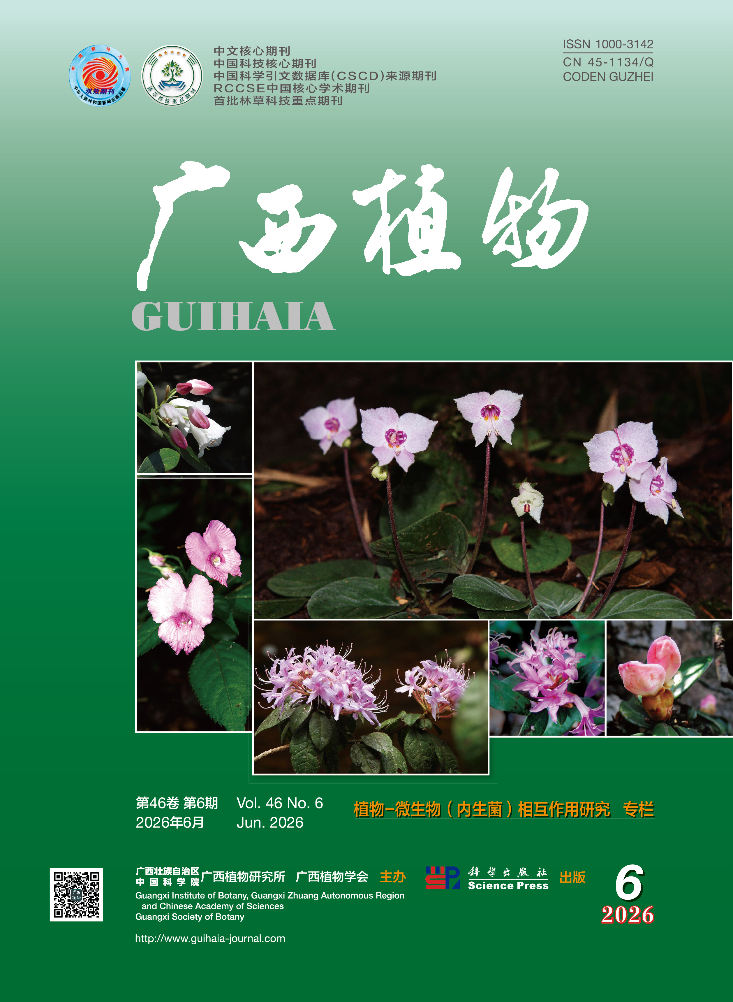
- 2026,No.6 Cover
- 2026,(6)
[Abstract](22) [PDF]()- 2026,No.6 Contents
- 2026,(6)
[Abstract](19) [PDF](2)Special Column: Research on Plant-Microorganism(Endophyte)Interactions
- AN Changrong1, LI Yun2, CHONG Huiying2, WEN Guangqin2, DUAN Ruyan1*
- Effects of inoculation methods of endophytic fungi on growth and physiological characteristics of blueberry seedlings
- To explore effective methods for inoculating blueberry with endophytic fungi, a pot experiment was conducted using one-year-old tissue-cultured blueberry seedlings and a strain of endophytic fungus(Talaromyces aculeatus). Five inoculation methods were designed, as root immersion in fungal suspension(F1), mixing fungal suspension with substrate(F2), inoculating with solid fungal blocks(F3), root immersion in fungal suspension after root pruning(F4), and irrigation with fungal suspension(F5). Seedlings immersed in sterile water without inoculation served as the control(CK). Root infection situation after inoculation was statistically analyzed, and determined some indexes that relate to growth and physiology. The results were as follow:(1)The root infection rates of blueberry were significantly different among the inoculation methods, the seedlings inoculated by F2 showed a higher root infection rate than those by other methods, and the root infection rate by F5 was the second highest. The root infection rates of treatments F1, F2, F3, F4 and F5 were 2.6, 3.7, 3.4, 3.2 and 3.6 times CK, respectively.(2)Except for leaf biomass and root biomass, all other growth parameters of blueberry seedlings inoculated with endophytic fungi were significantly higher than those of CK. Compared to the CK, the height increment, ground diameter increment, and total biomass of seedlings treated by five inoculation methods increased by 3.21%-30.47%, 16.37%-37.43%, and 9.69%-39.79%, respectively. The total root length, total root surface area, total root volume and root tip number increased by 38.63%-118.43%, 5.08%-94.89%, 11.97%-65.14%, and 28.90%-92.44%.(3)The physiological characteristic of blueberry seedlings by different inoculation methods showed significant differences, the maximum electron transport rate, minimum saturated light intensity, chlorophyll b content, and total chlorophyll content under the F3 were higher than other treatments, the chlorophyll a content and root activity showed higher with F2, while the CK had a higher initial slope than inoculation with endophytic fungi.(4)Based on the membership function analysis, the comprehensive evaluation result was F3 > F2 > F5 > F4 > F1 > CK. The inoculation method significantly affects the impact on the growth-promoting effect of endophytic fungi. The suitable inoculation methods are solid fungal blocks, mixing fungal suspension with substrate, and fungal suspension irrigation, which provide important insights for the efficient cultivation of endophytic blueberry seedlings.
- 2026,(6):919-930
[Abstract](22) [PDF](2)- LIU Yamin1, LING Qingyan1,2, ZHANG Beihong1,2*, ZHONG Qing3, LIU Xinli1, ZHAO Ruiqi1, HUANG Lina1, WANG Yanbo1,2, JIN Zhinong1,2
- Analysis on endophytic fungal community in Camphora tree species with citral chemotype
- To investigate the community diversity of endophytic fungi in three citral chemotype tree species in the Camphora, and to explore the diversity and structural differences of endophytic fungi in different tree species with the same chemotype and various tissue parts, this study analyzed 36 samples by high throughput sequencing method, a total of 2 207 878 sequences were obtained. The results were as follows:(1)The endophytic fungal communities of different tree species with the citral chemotype in the Camphora exhibited significant differences in species composition, and the endophytic fungal species composition showed organ preference and diversity in roots, stems, and leaves.(2)The ranking of the Chao and Ace indices was consistent, both showing Camphora officinarum (QCA)> C. bodinieri (QBO)> C. parthenoxylon (QPO), indicating that C. officinarum had the highest species richness of endophytic fungi, with 690 OTUs, while C. parthenoxylon had the lowest species richness, with 414 OTUs. In contrast, the Shannon diversity index showed the order QPO > QBO > QCA, and the Simpson diversity index exhibited the opposite trend. Thus, it could be concluded that the endophytic fungal community diversity of C. officinarum was the lowest.(3)Principal coordinate analysis(PCoA)based on Bray-Curtis distances revealed that the sample points of C. bodinieri and C. parthenoxylon were closely clustered, indicating a high similarity in their endophytic fungal community structures. In comparison, the sample points of these two species were relatively dispersed from those of C. officinarum, suggesting a lower similarity in endophytic fungal community structure between them and C. officinarum.(4)The three citral chemotype tree species of the Camphora all exhibited similar community structures of endophytic fungi in leaves and stems, but showed significant differences in the fungal communities between roots and leaves. The results indicate that the combined influence of host tree species and tissue heterogeneity shapes the endophytic fungal community, laying a foundation for exploring potential functional endophytic fungi in citral chemotype tree species of the Camphora and provide insights into the co-evolutionary relationship between endophytic fungal communities and host tree species and chemotypes.
- 2026,(6):931-942
[Abstract](20) [PDF](2)- YAN Zhongqi1, YANG Linsen2, YU Zhihe1, LIU Zhongyu1*
- Community structure and diversity of microorganisms from rhizosphere soil and root endosphere of two Cypripedium species in Shennongjia
- The growth and development of orchids are closely associated with microorganisms in rhizosphere soil and root endosphere. Characterizing orchid-associated microbial communities is of great significance for guiding artificial propagation, field conservation, and the exploration of potential functional microbial resources. To clarify the microbial community characteristics of endangered Cypripedium species in Shennongjia, wild Cypripedium henryi and C. japonicum were used as experimental materials. High-throughput sequencing was used to analyze the composition, diversity, and potential functions of fungal and bacterial communities in rhizosphere soil and root endosphere at the flowering and fruiting stages. The results were as follows:(1)A total of 14 fungal phyla and 38 bacterial phyla were detected in rhizosphere soil and root endosphere of the two Cypripedium species. The dominant fungal phyla were Ascomycota, Basidiomycota, and Mortierellomycota, and the dominant bacterial phyla were Proteobacteria, Acidobacteriota, and Actinobacteria.(2)The richness and evenness of fungal and bacterial communities in rhizosphere soil were generally higher than those in root endosphere. Fungal richness in rhizosphere soil increased significantly from the flowering stage to the fruiting stage, whereas bacterial richness in root endosphere decreased significantly.(3)Ecological niche explained a relatively high proportion of the differences in microbial community structure between rhizosphere soil and root endosphere of the two Cypripedium species, accounting for 19.49% and 60.20% of the variation in fungal and bacterial communities, respectively.(4)Developmental stage affected the abundance changes of some microbial taxa and differentiation of community functions. From the flowering stage to the fruiting stage, the relative abundance of 13 fungal genera in rhizosphere soil and 20 bacterial genera in root endosphere changed significantly.(5)The functional composition of fungal communities differed markedly between rhizosphere soil and root endosphere, whereas the functional profiles of bacterial communities were relatively stable. Competitive and cooperative interactions coexisted in rhizosphere soil microbial communities, whereas positive correlations dominated in root-endosphere microbial communities. This study preliminarily clarifies the composition characteristics of fungal and bacterial communities in rhizosphere soil and root endosphere of two Cypripedium species in Shennongjia, as well as their associations with ecological niche, developmental stage, and host species. These findings provide foundational data for screening symbiotic microorganisms, artificial propagation, and population conservation of Cypripedium plants.
- 2026,(6):943-956
[Abstract](20) [PDF](2)- GUO Zhenghu1,2,3, WANG Jie1,3, TAN Xiaoming1,2,3,4*, YAO Chun1,2, LI Liangbo1,2,4, QIN Qinjuan5, HUANG Rongshao1,2,4
- Effects of endophytic fungus Subsessila turbinata RG170 on physiological characteristics and secondary metabolite in Cinnamomum cassia seedlings
- To investigate the effects of the endophytic fungus RG170(Subsessila turbinata)on the physiological characteristics and secondary metabolite accumulation of Cinnamomum cassia seedlings, the endophytic fungus RG170, isolated from healthy leaves of C. cassia, was used as the research subject. The plant growth-promoting potential was evaluated through in vitro assays assessing phosphate solubilization, potassium solubilization, and nitrogen fixation, as well as through a sterile co-culture system. Pot experiments were further conducted to verify its actual effects on C. cassia growth and metabolism. The results were as follows:(1)Under sterile co-culture conditions, inoculation with RG170 significantly promoted the growth of C. cassia seedlings. Compared with the control, plant height, fresh weight, number of lateral roots, and leaf area increased by 15.4%, 54.2%, 40.1%, and 29.5%, respectively, with all parameters except plant height showing significant differences.(2)In pot experiments, inoculation with RG170 significantly increased the dry biomass of the underground parts of C. cassia seedlings.(3)Compared with the control, the RG170-inoculated seedlings exhibited significantly higher levels of soluble sugars and soluble proteins in leaves, with increases of 51.9% and 63.8%, respectively, while the activities of superoxide dismutase(SOD)and catalase(CAT)were significantly enhanced by 66.5% and 279.0%, respectively.(4)Inoculation also significantly increased total flavonoid and total phenolic contents in leaves by 115.3% and 113.9%, respectively; the contents of cinnamyl alcohol, cinnamic acid, and cinnamaldehyde in both leaves and stem bark were significantly elevated, with cinnamaldehyde content increasing by 35.5% and 68.8%, respectively. In conclusion, inoculation with the RG170 strain, which possesses multiple growth-promoting traits, not only promotes the growth and enhances the physiological activity of C. cassia seedlings but also facilitates the accumulation of key secondary metabolites, such as total flavonoids and cinnamaldehyde. This study provides a basis for the development of microbial inoculants for C. cassia.
- 2026,(6):957-967
[Abstract](22) [PDF](3)植物生态学
- YU Ling1,3, HE Wen2,3*, LIU Peng1, LIU Siqin1, LING Jiaxuan1, LI Mengmeng1, LI Ning4, JIA Bin5
- Spatiotemporal variations of forest NDVI in Guangxi and its sensitivity to drought
- To investigate the sensitivity of forests in Guangxi to drought and to reveal the characteristics of stability, resistance, and resilience of forest ecosystems under drought stress, standardized precipitation evapotranspiration index(SPEI)and normalized difference vegetation index(NDVI)were used to analyze stability, resistance, and resilience of forests in Guangxi during 2010-2022. The results were as follows:(1)During the study period, drought conditions in the Guangxi forest region exhibited an overall pattern characterized by an initial decreasing trend followed by an increasing trend, indicating a shift from relatively wetter conditions to increased drought stress in recent years.(2)Over the past decade, forest NDVI in Guangxi increased at a rate of 0.06 a-1, with greater increases observed in karst region than in non-karst region, and in planted forest than in natural forest, suggesting that vegetation growth dynamics differed significantly across regions and forest types.(3)The spatial pattern of NDVI stability was similar to that of resistance under drought conditions, whereas resilience exhibited an opposite spatial pattern, reflecting a trade-off between the ability of forests to resist drought disturbances and their capacity to recover after disturbance.(4)The stability and resistance of forest NDVI in non-karst region were higher than those in karst region, while the resilience was greater in karst region; in addition, natural forest exhibited higher stability, resistance, and resilience than planted forest. Overall, these results indicate that the response of forest NDVI to drought in Guangxi exhibits pronounced regional and forest-type differences, and provide a scientific basis for adaptive forest management and ecosystem restoration under drought conditions.
- 2026,(6):968-977
[Abstract](22) [PDF](3)- HU Jiaxue1, WU Qian1,3, LI Yin1, LI Zongyan1*, RUAN Yuehong1, YANG Jianmin1, NIAN Yaoping2
- Comparison of nitrogen use efficiency among different ecotypes of Paphiopedilum micranthum and their carbon-nitrogen allocation strategies
- To reveal the nitrogen adaptation mechanisms of different Paphiopedilum micranthum ecotypes in karst habitats, this study focused on three ecotypes(lithophytic type, scrub terrestrial type, and understory terrestrial type)co-occurring in a limestone area in southeastern Yunnan. We aimed to compare their nitrogen uptake and utilization efficiencies and elucidate their ecological adaptation strategies from the perspective of carbon-nitrogen allocation. By measuring soil available nitrogen content, and analyzing plant N phenotypes along with the activities of key N metabolism enzymes. We compared inter-ecotype differences in N accumulation and utilization efficiency, followed by correlation analysis of influencing factors. The results were as follows:(1)While no significant difference was observed in nitrogen uptake efficiency(NUpE), nitrogen use efficiency(NUE)differed significantly among ecotypes(P<0.05). The lithophytic type [(2.83±0.14)g·mg-1] and the understory terrestrial type [(2.83±0.05)g·mg-1)exhibited significantly higher NUE than the scrub terrestrial type [(2.64±0.22)g·mg-1).(2)Divergent adaptive characteristics were formed: the understory terrestrial type had significantly greater total root length [(64.80±9.86)cm)and root-to-shoot ratio(0.95±0.22)than the lithophytic type [total root length:(50.26±17.50)cm; root-to-shoot ratio: 0.72±0.17]. In contrast, the lithophytic type displayed the highest nitrogen uptake per unit root length, significantly surpassing the understory terrestrial type(P<0.05). The scrub terrestrial type demonstrated a strong nitrate assimilation capacity, possessing the highest nitrate reductase activity [(49.37±1.08)μg NO2-·g-1 FW·h-1] and leaf N concentration.(3)Carbon-nitrogen allocation strategies differed markedly: the lithophytic type had the highest whole-plant carbon-to-nitrogen ratio(C/N), whereas the scrub terrestrial type exhibited the greatest C and N accumulations but the lowest C/N. The understory terrestrial type generally displayed intermediate values for these traits.(4)The correlation analysis showed that, nitrogen use efficiency was significantly positively correlated with total root length, root carbon accumulation, and leaf C/N, and significantly negatively correlated with soil ammonium nitrogen content. In conclusion, the different ecotypes develop complementary nitrogen adaptation mechanisms by regulating root architecture and carbon-nitrogen allocation. The lithophytic type achieved the highest nitrogen use efficiency through more nitrogen uptake per unit root length and a resource allocation strategy characterized by a higher C/N. By comparing the differences in nitrogen uptake/use efficiency and N metabolism physiology among ecotypes, this study elucidates their ecological adaptation mechanisms from the perspective of carbon-nitrogen allocation, and provides a theoretical basis for the scientific conservation and cultivation of this species.
- 2026,(6):978-990
[Abstract](20) [PDF](1)- LIANG Yuying1, HE Qinxia1, LI Jiyin1, MA Hailun1, HUANG Haimei1, ZHENG Jiamin1, MING Angang2,3, WANG Qingling2,3, HUANG Xueman1,3, YOU Yeming1,3*
- Effects of introducing nitrogen-fixing tree species on soil-microbial ecological stoichiometric characteristics in karst areas
- Ecological stoichiometry primarily investigates the proportional relationships of chemical elements between organisms and their environment within ecosystems. It serves as a foundation for understanding life activities and ecosystem functions. The study of soil-microbial biomass and their ecological stoichiometric characteristics of different types of tree species in karst areas is crucial for scientifically assessing the effectiveness of various tree types in improving soil nutrient conditions and optimizing tree species configuration strategies. This study was conducted in Daqingshan Stone Mountain Arboretum at the Experimental Center of Tropical Forestry, Chinese Academy of Forestry. Five nitrogen-fixing tree species(N-fixer)and seven non-nitrogen-fixing tree species(non-N-fixer)were taken as study objects. The research investigated the response patterns of soil-microbial carbon(C), nitrogen(N)and phosphorus(P)contents to N-fixer and non-N-fixer in karst ecosystems. It also analyzed ecological stoichiometric ratios, microbial quotient(qMBC, qMBN and qMBP), and soil-microbialstoichiometric imbalance(Cimb:Nimb, Cimb:Pimb and Nimb:Pimb). The results were as follows:(1)The total nitrogen(Nsoil)content and total phosphorus(Psoil)content of the soil in N-fixer tree species were significantly higher than that in non-N-fixer, but Csoil:Nsoil was significantly lower than those in non-nitrogen-fixer.(2)The contents of microbial biomass carbon(MBC), microbial biomass nitrogen(MBN)and microbial biomass phosphorus(MBP)were significantly higher in N-fixer than in non-N-fixer. In contrast, the ratios of MBC:MBP and MBN:MBP were significantly lower in N-fixer than in non-N-fixer. No significant differences were observed between N-fixer and non-N-fixer for Cimb:Nimb, Cimb:Pimb and Nimb:Pimb, indicating that they were characterized by a certain degree of internal stability.(3)qMBC of N-fixer was significantly larger than that of non-N-fixer, while qMBN and qMBP showed no significant difference between these two types of tree species. The RDA results showed that Csoil:Psoil, MBN:MBP and Csoil:Nsoil were the key factors influencing soil microbial quotient. This study indicates that compared with non-N-fixer, N-fixer has significant advantages in improving soil nutrient status and alleviating soil P limitation in karst areas, which provides an important scientific basis for the selection of tree species in the process of ecological restoration.
- 2026,(6):991-1002
[Abstract](22) [PDF](2)- ZHANG Yaoyao1, REN Qifei2,3*, FU Hangqin1, CHEN Yunfei2,3
- Effects of formula fertilization on seedling growth and ecological stoichiometric characteristics of Davidia involucrata
- To explore the optimal fertilizing amount of nitrogen(N), phosphorus(P)and potassium(K)for the growth of Davidia involucrata seedlings, a pot control experiment was conducted with one-year-old seedlings as research object and yellow brown soil as the substrate whose basic physical and chemical properties were determined. The “3414” soil testing and formulated fertilization method was adopted to set three factors(N, P, K)with four levels(0, 1, 2, 3), with a total of 14 treatments in all(T1-T14). The seedling growth and ecological stoichiometric characteristics of leaves and roots under different treatments were determined, and the inter-treatment differences and correlation relationships of each index were statistically analyzed. The results were as follows:(1)The carbon(C)content in leaves and roots of D. involucrata seedlings showed relatively small differences among all treatments. The N and P contents showed significant differences(P<0.05), and they were positively correlated with the application levels of N and P fertilizers, respectively.(2)The C:N and N:P in leaves and roots under T2(N0P2K2)were unbalanced, which limited the growth of D. involucrata seedlings. The N:P ratios in leaves and roots among all treatments ranged from 4.86 to 13.65, all lower than 14, indicating the seedling growth was limited by N.(3)The leaf mass per area, total biomass, plant height increment and ground diameter increment were the lowest under T11(N3P2K2), which were 6.78%, 32.88%, 18.91% and 6.82% lower than those under T1(N0P0K0)respectively. T13(N1P2K1)had the most significant growth-promoting effect, with the above indicators being 12.39%, 82.37%, 192.08% and 120.45% higher than those under T1 respectively.(4)Correlation analysis showed that leaf mass per area was significantly positively correlated with root N content and N:P of leaves and roots(P<0.05), and total biomass was significantly positively correlated with root C content(P<0.05), indicating a certain correlation between growth traits and ecological stoichiometric characteristics.(5)Multiple comparison results showed that N had significant effects on all growth traits, as well as root C and N contents and leaf N and P contents(P<0.05). P had significant effects on leaf mass per area, plant height increment, ground diameter increment and P contents of leaves and roots(P<0.05). K had a significant effect on leaf mass per area, plant height increment and ground diameter increment(P<0.05). In conclusion, balanced application of N, P and K should be emphasized for potted D. involucrata seedlings, and excessive application of N fertilizer(7.95 g·plant-1)should be avoided due to its inhibitory effect on seedling growth. T13(2.65 g N, 1.00 g P2O5, and 1.50 g K2O)is the optimal fertilization scheme for the growth of D. involucrata seedlings. The precise fertilization model constructed in this study provides core technical support for the population propagation and natural reintroduction of D. involucrata, and contributes to the conservation of this endangered species resource.
- 2026,(6):1003-1015
[Abstract](18) [PDF](2)- HU Xiaoxue1,2, GUO Jing1,2, NONG Junqing2, LI Zailiu1*, XU Zhenguo2*
- Characteristics of C, N, and P stoichiometric dynamics during flowering period of Phyllostachys edulis
- Ecological stoichiometry is a discipline that elucidates how plants regulate nutrient allocation and utilization across critical life-history stages. To explore the stoichiometric adaptation mechanisms and nutrient resource allocation strategies of bamboo plants, using naturally flowering P. edulis in northern Guilin, Guangxi, China as the study material, we quantified carbon(C), nitrogen(N), and phosphorus(P)contents, along with their stoichiometric ratios(C:N, C:P, N:P)across seven organ types [culm, branch, leaf/flower spike, culm stump, culm root, rhizome, and rhizome root] during four flowering periods: pre-flowering, initial flowering, peak flowering, and late flowering. The objective was to elucidate the underlying stoichiometric adaptation mechanisms and resource reallocation strategies that underpin reproductive investment in bamboo. The results were as follows:(1)The C, N, and P contents in different organs exhibited significant differences and temporal dynamics across all four periods. Branches consistently exhibited the highest C content; leaf/flower spike N peaked during pre-flowering, whereas P peaked at peak flowering; culm P, branch C, and leaf N declined monotonically throughout flowering; rhizome C and N showed a unimodal pattern(decreasing then increasing); and P in branches, culm roots, and rhizome roots followed an inverse unimodal trajectory(increasing then decreasing).(2)Stoichiometric ratios displayed distinct spatiotemporal patterns. C:N reached its maximum in culms during initial flowering; C:P peaked in rhizome roots at late flowering; and N:P was the highest in rhizomes during pre-flowering.(3)Significant(P < 0.05)or highly significant(P < 0.01)correlations were observed among C, N, and P contents and ratios across organs within each period, indicating tightly coordinated, stage-dependent nutrient partitioning. In conclusion, during the flowering period of P. edulis, vegetative organs such as branches accumulate C to maintain homeostasis, while leaves/flower spikes accumulate N and P to support metabolic processes. This overall pattern reflects an ecological adaptation strategy characterized by “C homeostasis, P limitation, and N redistribution” ensuring reproductive demands.
- 2026,(6):1016-1026
[Abstract](20) [PDF](2)- LI Shengfan1, LÜ Shi2, ZHENG Renhao1, LIN Hui2, DU Hongwei1, YAN Huihui2, ZHOU Ran1, WANG Ziyi1, LIN Dongmei2, LIN Zhanxi2, LIU Fengshan2*
- Comparative analysis of leaf spectral characteristics of different Juncao species based on visible-near infrared spectroscopy and machine learning
- Aiming to address the challenges of morphological similarity among different species of Juncao and the low efficiency of traditional identification methods, this study employs spectral feature analysis and machine learning modeling to achieve efficient identification and classification of eight Juncao species. Spectral reflectance data of leaf samples from eight species, including Cenchrus fungigraminus, Penmisetum purpureum, P. purpureum cv. Laimu-1, P. purpureum cv. Red, P. alopecuroides, P. glaucum × purpureum, P. americanum, and Saccharum officinarum cv. PNG, were collected within the 400-900 nm wavelength range. By processing the original spectra along with their reciprocal taking logarithm transformations, the first derivatives, and the second derivatives, spectral characteristics were analyzed, and multiple vegetation indices were extracted. Based on six types of feature sets — original spectra, reciprocal taking logarithm transformation, the first derivative, the second derivative, three-edge parameters, and vegetation indices — twenty graded feature combinations were constructed. Classification models were developed using support vector machine(SVM)and random forest(RF)algorithms, with model accuracy evaluated accordingly. The results were as follows:(1)The eight Juncao species exhibited typical vegetation spectral characteristics in the visible light region, with reflectance rising sharply in the red-edge region(700-750 nm).(2)Different spectral processing methods significantly amplified inter-species spectral differences in specific bands, such as reciprocal taking logarithm transformation within 570-650 nm, the first derivatives around 730 nm, and the second derivatives in the 670-760 nm range. The red-edge amplitude, red-edge area, and simple ratio(SR)vegetation index demonstrated the strongest discriminative power among species.(3)Model performance indicated that the SVM algorithm generally outperformed RF. The combination of “original spectra + three-edge parameters” achieved the highest accuracy of 70.56% under SVM, which was 14.03% higher than the same combination under RF. The optimal performance for RF was observed with the “original spectra + reciprocal taking logarithm transformation” combination, though it showed limited adaptability to high-dimensional features. Integrating spectral features from visible and near-infrared bands with spectral transformation techniques and vegetation indices, coupled with the SVM algorithm, provides an effective theoretical foundation and technical support for the rapid and accurate identification and classification of Juncao species.
- 2026,(6):1027-1045
[Abstract](24) [PDF](2)- LING Lei1,2, REN Zengcao1,2, ZONG Wenzhen1, DING Aiqiang1,2, CHEN Zhengni1,2*
- Effects of stand age and density on carbon storage of Picea asperata plantations in the Taohe River basin
- In the context of global carbon neutrality and regional ecological security requirements, the optimization of the carbon sink function of Picea asperata plantations in the Taohe River basin has become increasingly crucial. To explore the distribution characteristics and influencing factors of its carbon storage, this study focused on P. asperata plantations within the Taohe River basin, establishing plots with varying stand ages(young and middle forests)and densities(925-2 800 trees·hm-2). It systematically measured and comprehensively evaluated the carbon storage and distribution characteristics of different organs(trunk, branch, leaf, bark, root)within the tree layer, the litter layer, and distinct soil horizons(0-10 cm, 10-20 cm, 20-40 cm, 40-60 cm, 60-80 cm). It provided an in-depth analysis of the mechanisms by which stand age and density influenced carbon storage. The results were as follows:(1)The total carbon storage of P. asperata plantation ecosystem in the Taohe River basin ranged between 336.25 to 512.39 t·hm-2. The overall pattern exhibited a hierarchy of carbon storage: soil layer > tree layer > litter layer.(2)The soil layer constituted the most fundamental stable carbon reservoir within ecosystems, holding carbon storage ranging from 256.89 to 300.45 t·hm-2. This accounted for 69.90% of total carbon storage, with over 60% of soil carbon concentrated within the 0-40 cm soil layer.(3)The carbon storage in the tree layer ranged between 43.12 to 190.02 t·hm-2, exhibiting pronounced spatio-temporal heterogeneity alongside a significant ‘mid-mature forest carbon accumulation peak'. Carbon was preferentially allocated to the trunk(22.46%-49.42%)and root(19.39%-29.35%)among the various organs. Its distribution strategy was density-regulated: biomass in high-density stands concentrated in the trunk, while in medium-to-low-density stands, all organs grew more evenly, resulting in higher carbon sequestration efficiency.(4)The carbon storage in the litter layer ranged from 12.53 to 21.92 t·hm-2, accounting for 3.58% to 4.30% of the total carbon storage. However, its carbon content was remarkably high at 306.04 to 389.32 g·kg-1, making it a pivotal hub connecting above-ground and below-ground carbon flows. This study confirms that stand age and density are the core factors driving carbon accumulation and distribution within P. asperata plantations ecosystems. By implementing sustainable management strategies centred on density regulation, the carbon sink potential of P. asperata plantations in this region can be effectively harnessed. The results provide crucial theoretical basis and practical guidance for optimizing the carbon sink function of plantations in the Taohe River basin, safeguarding ecosystem, and achieving regional carbon neutrality objectives.
- 2026,(6):1046-1055
[Abstract](24) [PDF](2)- ZHANG Xinran, ZHAO Suya, WANG Yifan, ZHENG Yuxin, ZHANG Chan*
- Reproductive strategies in alpine extreme environments: the plastic flower longevity and fly pollination in Gentiana aristata
- Gentiana aristata, a typical alpine species of the Gentiana genus, serves as an ideal research material for analyzing species adaptation mechanisms to extreme environments, developing distinctive medicinal bioactive compounds, and breeding cold-resistant ornamental germplasm. To uncover the pollination biology characteristics of G. aristata and its reproductive adaptation strategies to alpine extreme environments, this study employed a combination of field observations and controlled experiments to systematically investigate its reproductive traits, including flowering dynamics, pollen viability, stigma receptivity, pollen/ovule ratio(P/O), breeding system, and composition of pollinating insects. The results were as follows:(1)G. aristata exhibited herkogamy and incomplete dichogamy. The longevity of individual flower of G. aristata was(5.97±0.19)d, which significantly extended to(16.13±0.78)d under insect-excluded conditions, representing an adaptive strategy to the variable pollination environment in alpine regions.(2)Both pollen viability and stigma receptivity of G. aristata were maintained for approximately 4 d, with pollen viability peaking on the second day after anthesis and stigma receptivity reaching its maximum on the second day after the stigma fully opened.(3)The results of breeding system showed the fruit setting in bagging without emasculation flowers was primarily attributed to autonomous selfing via temporary corolla closure and assisted selfing by small insects like thrips(Thripidae spp.)and ants(Formica spp.). The fruit setting rates and seed setting rates were low and showed no significant difference between those flowers under natural pollination after emasculation and bagging without emasculation, indicating that both selfing and outcrossing have similar reproductive success, but both were lower than that under natural pollination.(4)The main pollinator of Gentiana aristata was Delia platura, which accounted for 55.17% of all flower visits, with a visit frequency of(0.24±0.04)times·flower-1·h-1. In addition to D. platura, other pollinators included various flies and beetles. In summary, Gentiana aristata exhibits high plasticity in flower longevity and possesses a mixed mating system combining selfing and outcrossing. Its main pollinators are abundant flies that show relatively high floral visitation frequency. These traits collectively represent a key reproductive strategy for adapting to the unpredictability of alpine pollination environments. The findings provide critical theoretical support for the conservation of G. aristata germplasm resources and its medicinal development.
- 2026,(6):1056-1065
[Abstract](22) [PDF](2)- MAIMAITI Mierkamili1,2, LIU Jingwen1, MA Xinhui1, ZHANG Hanrui1, TANG Jia1, TIAN Zhongping3, ZHANG Kai1,2*
- Population structure and dynamic characteristics of the natural Caragana turfanensis in the Tomur Peak National Nature Reserve
- Caragana turfanensis is a near-threatened plant endemic to Tianshan Mountains of China. To clarify the survival status and developmental trends of its populations, this study investigated the natural populations of C. turfanensis in the Tomur Peak National Nature Reserve. Using quadrat surveys and population statistical methods, we compiled a static life table, fitted survival curves, and conducted a systematic assessment combined with population viability and dynamic quantitative indices. The results were as follows:(1)The population exhibited an “inverted J-shaped” age structure, with a high proportion of seedlings and young individuals, indicating an expanding growth pattern, yet the age-class distribution was uneven.(2)The survival curve conformed to the B3 subtype of the Deevey Type II curve, characterized by extremely high early mortality(up to 90.5% in age class I), reflecting strong environmental filtering.(3)Survival analysis revealed that the survival rate declined sharply with increasing age class(dropping to only 0.9% by age class III), with cumulative mortality reaching 99.1% by age class III, indicating increasing mortality in older classes.(4)Population dynamic index analysis indicated that the dynamic indices of all age classes of C. turfanensis were positive, reflecting an overall increasing population trend. However, the V'pi value(3.14%)was close to zero, implying extremely slow growth under natural disturbances. The maximum risk value of random disturbance for the population(Pmax=0.04%)demonstrated weak resistance to environmental interference and high ecological vulnerability. In conclusion, although C. turfanensis populations possess growth potential, the extremely high early mortality causes a severe “seedling bottleneck”, resulting in poor population stability. It is recommended to implement measures such as microhabitat improvement, establishment of protected micro-reserves, and enhancement of artificial assisted regeneration to break through the early survival bottleneck and promote population recovery and stability.
- 2026,(6):1066-1075
[Abstract](20) [PDF](2)- QIN Zhiwei1,2, MENG Jian1, XU Zhanyong1, LUO Weisheng1, LU Feng1, LIU Xionghui 2, TANG Qiuyue2, QIN Kun3, TAN Bizeng1*
- Water-holding and water-losing characteristics of litter in different forest types in Dayaoshan Reserve, Guangxi
- The water-holding and water-losing characteristics of forest litter are critical components regulating hydrological processes in forest ecosystems. However, the quantitative characterization and mechanistic analysis of the hydrological functions of the litter layer across different forest types remain largely inadequate to date. To clarify the water-holding and water-losing characteristics of litter in major forest types in the Dayaoshan National Nature Reserve, Guangxi, this study employed a combination of field plot surveys and laboratory-controlled experiments to determine the litter standing crop, dynamic water-holding processes, and dynamic water-losing processes of three typical forest types in the reserve, namely evergreen broadleaf forest(EBF), mixed coniferous-broadleaf forest(MCBF), and montane elfin forest(MEF). Furthermore, their water conservation capacity was evaluated. The results were as follows:(1)The total litter accumulation of three forest types ranged from 2.28 to 5.33 t·hm-2, with the order of EBF> MCBF> MEF.(2)The relationship between water-holding capacity(Q)and immersion time(t)followed the logarithmic function [Q=aln(t)+b], and the relationship between water-absorption rate(V)and immersion time(t)followed the power function(V=ktn); there was a significant logarithmic relationship between water-losing capacity and seasoning time(R2>0.95, P<0.05), whereas a significant power function relationship was found between water-losing rate and seasoning time(R2>0.95, P<0.05).(3)The effective interception rate ranged from 49.46% to 116.13%, and the effective interception capacity ranged from 0.41 to 3.09 t·hm-2. Among them, the effective interception capacity of EBF and MCBF was significantly higher than that of MEF(P<0.05). In conclusion, there were significant differences in the hydrological regulation efficiency of litter among different forest types in the Dayaoshan Reserve, Guangxi. EBF not only had the highest rainfall interception capacity but also exhibited better coordination between water-holding and water-release. The results of this study can provide empirical basis and parameter support for the accurate evaluation of water conservation functions, quantification of ecological benefits, and protection and management decisions of subtropical montane forests.
- 2026,(6):1076-1086
[Abstract](20) [PDF](1)- PAN Yinxixue1, WANG Xuezhao2,3,4, LI Xiaona2,4*, ZHANG Lin1, WANG Leiguang5
- Impacts of climate change on the potential ecologically suitable plantation areas of Macadamia sp. in China
- Climate change has exerted profound impacts on the distribution patterns of plant species. Macadamia sp., a tropical to subtropical montane plant, possesses considerable economic and ecological significances. Understanding its potential ecologically suitable areas under future climate change scenarios is of great importance for promoting sustainable economic development and soil and water conservation in mountainous regions of China. This study integrated the MaxEnt model with ArcGIS technology to analyze current distribution data of Macadamia sp. in China, along with environmental variables including climatic, topographic, and soil factors, etc. Key environmental determinants influencing its distribution were identified, and the potential distribution patterns of suitable areas were projected for four future time periods: the 2040s, 2060s, 2080s, and 2100s. The results were as follows:(1)The MaxEnt model demonstrated high accuracy in simulating the potential geographic distribution of Macadamia sp. in China.(2)Annual mean temperature, annual mean precipitation, isothermality, and temperature annual range were identified as the most influential environmental variables shaping its distribution.(3)Across all climate scenarios, Yunnan Province consistently exhibited the largest distribution area of suitable area, followed by the southeastern coastal provinces such as Guangdong and Guangxi.(4)Under all three future climate scenarios(SSP1-2.6、SSP2-4.5、SSP5-8.5), the total area of suitable areas gradually decreased over time, accompanied by increasing fragmentation, while the spatial shifts of the overall centroid remained minimal. These findings provide a comprehensive understanding of the potential ecological distribution patterns, and offers a scientific basis for the development of large-scale cultivation strategies.
- 2026,(6):1087-1100
[Abstract](18) [PDF](2)
